Microscopes, a quick look back, then zoom to the present...
It’s easy to take the existence of many tools in the conservation studio for granted, no matter how simple they may be in design, but the story and development of the microscope is probably one that will continue ad infinatum.
Just take me to the techy bit
There are records which speak of the use of water for magnification as far back as 4000 years ago in both ancient China and Greece. 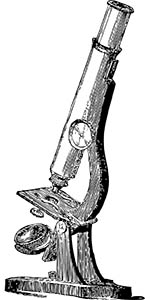 The use of curved lenses for magnification came significantly more recently in human history, around the 14th century, in the form of spectacles. It’s not too much of a leap for magnifying glasses to be invented, but It took another couple of hundred years for a microscope, as we’d know it, to appear. Many claim to have invented the ‘microscope’, and it seems likely it happened between 1590 and 1620, with ‘the’ Galileo as one of those with his name ‘in the hat’.
It’s easy to understand the fascination with the ‘new world’ microscopes offered a window into, biological analysis had never before been so detailed. Illustrations of this new world produced by Robert Hooke, a British scientist (as well as being a map maker, architect, astronomer) offered others a glimpse of details never seen before in his book Micrographia, published in 1665. You can read more about ‘Micrographia’ on the British Library website here.
In 1676 Antonie van Leeuwenhoek a dutch scientist, who had conversed regularly with Robert Hooke after the publication of Micrographia, reported the discovery of micro-organisms, he called them ‘animalcules’, we now know these as bacteria. Van Leeuwenhoek had managed to achieve magnification levels of up to 300x with his simple single lens design.
Lens refinement was the main development in microscope technology for the next few centuries. It took the advent of electrical illumination in the late 19th century to progress the light microscope’s development to near its fullest potential.
That’s history, what’s new?
Of course, fast-forwarding to the present, things have moved on substantially with digital technology providing many more options.
Some features that you will see on our digital microscope range are the following:
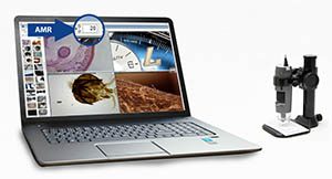
AMR -
(Automatic Magnification Reading) Displays the current level of magnification on-screen. Each image captured or filmed by the microscope displays the level of magnification automatically. This means that there is no need to manually annotate images or files, and that no images are saved without annotation. This feature is compatible with Windows only.
See Microscopes with the AMR function here.
EDOF -
(Extended Depth of Field) Combines images at different focus levels for overall clarity on uneven surfaces, also known as ‘Focus stacking’. When working at high magnification there is a limited depth of field, meaning some of the detail of a 3D object can be out of focus. The software captures the images with a single click and combines them automatically onto one composite image. Manual mode is also available which allows users to select the images they would like to use in the composite image. This is a valuable feature when working on any object with an uneven surface or multiple visible layers, preventing loss of clarity and the need to capture multiple images. This feature is compatible with Windows only. See Microscopes with the EDOF function here.
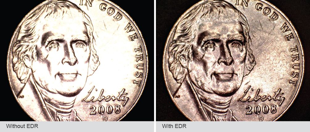
EDR –
(Extended Dynamic Range) Increases the dynamic range by combining images taken at different exposure levels into one image, with only one click. When viewing highly reflective objects like metals, glass, ceramics, stone, or those with uneven lighting, details can be lost or obscured (i.e. in a reflection or a shadow). The software automatically selects the best combination of images to create a composite image with the best possible lighting. This feature is compatible with Windows only. See Microscopes with the EDR function here.
FLC –
(Flexible LED Control) With 4-quadrants switching capability and 6-levels luminance adjustability, the FLC provides greater control of the built-in LED illumination. This is particularly useful when capturing the texture of a surface, for instance viewing stroke direction in a painting, or abrasions to a polished surface. This feature is compatible with Windows only.
See Microscopes with the FLC function here.
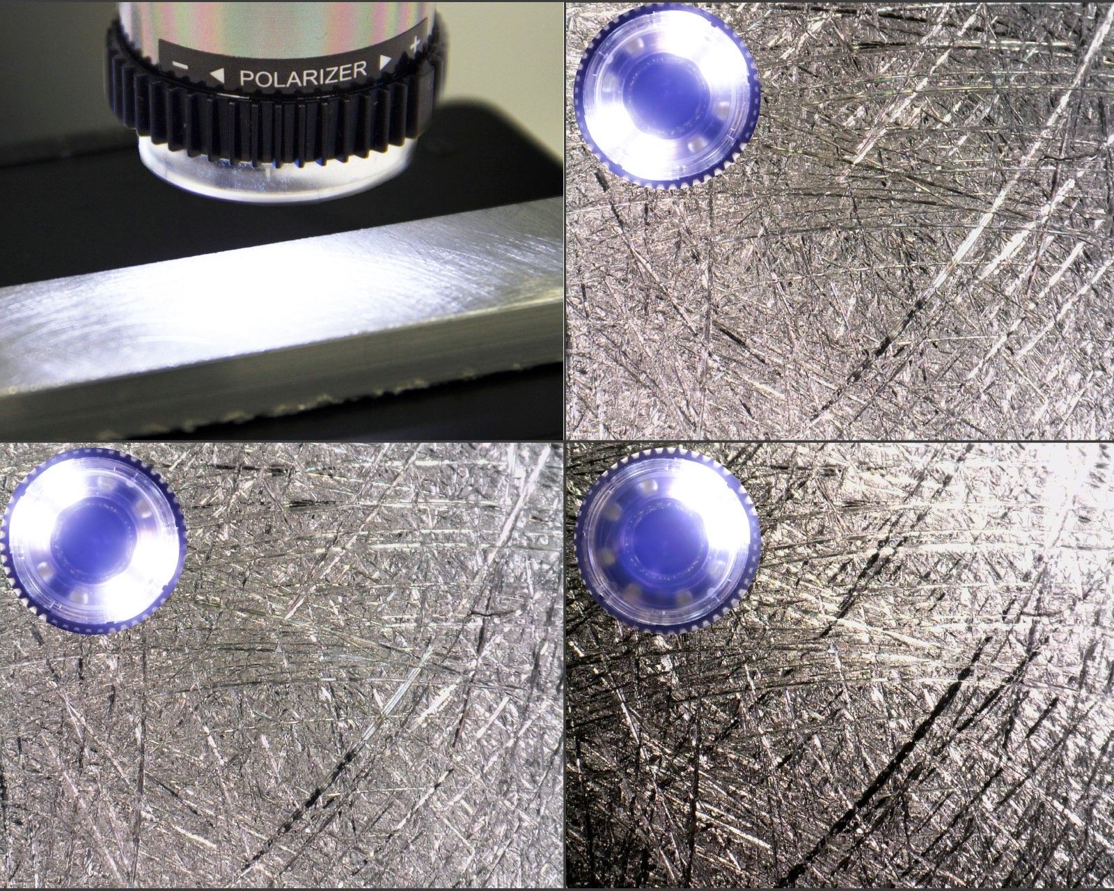
Exchangeable Caps
Exchangeable caps provide extra flexibility in use. The extra caps offer further options for lighting and magnification. The closed cap is particularly useful in conservation cleaning as it prevents dust or liquid from touching the lens.
Measurement and Annotation
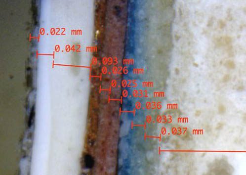
As part of the included software, all of our digital microscopes offer a measurement and annotation function for captured images.
You can read more about the software here.
Working Distance
Microscope working distances are fixed, with the highest levels of magnification available at the closest focusable distance from the object. Standard working distance microscopes focus from ‘almost touching’, to around 50mm away from the object. Long working distance models are able to focus from around 30-40mm up to 230mm away from an object. When selecting a microscope for the purposes of cleaning or detailed repair, a long working distance model is recommended, enabling plenty of space in which to work between the microscope and object. You can view each microscope’s working distance in the ‘Specification’ tab.
Polarising
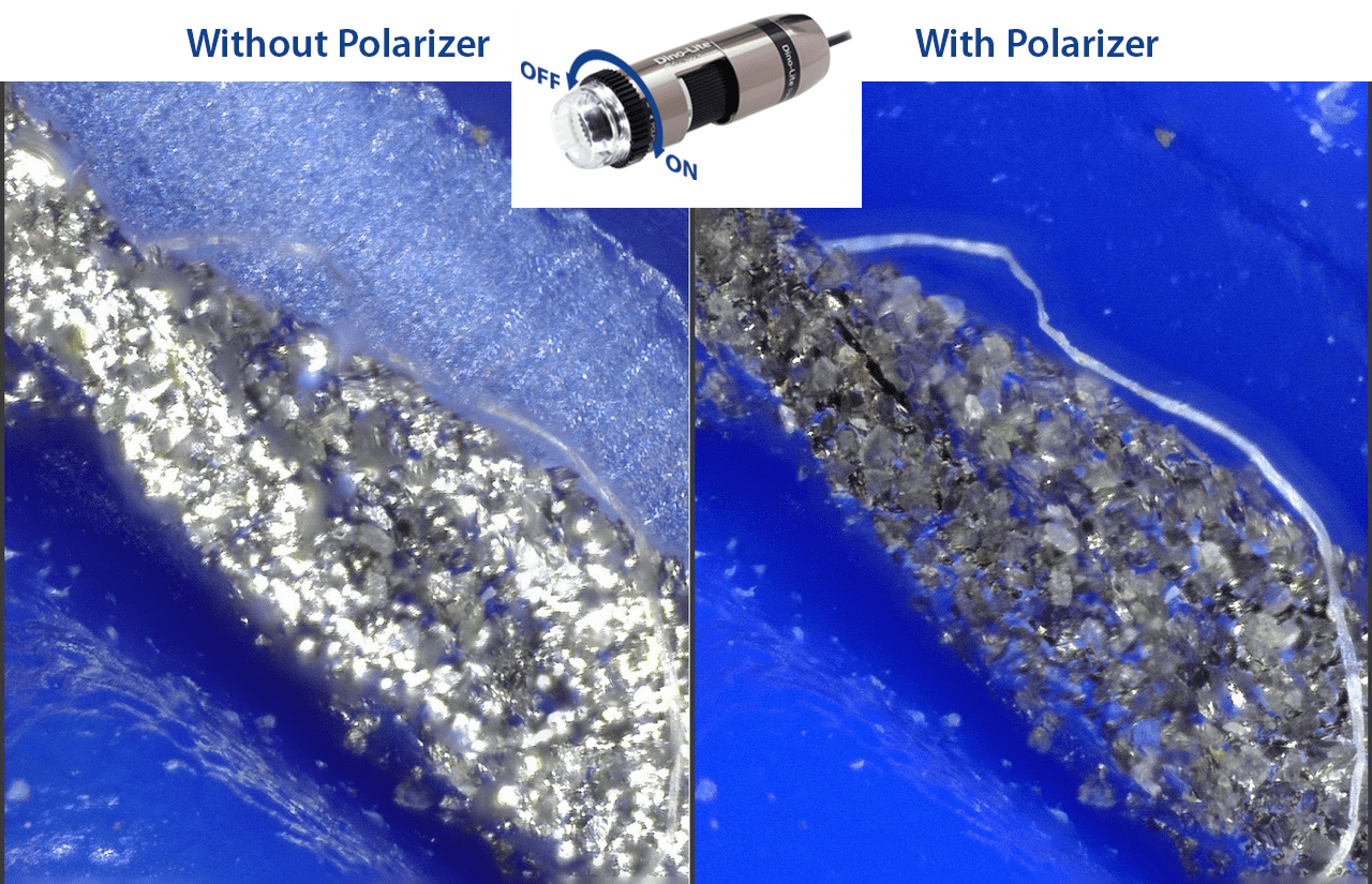
Microscopes which feature the polarising filter are especially useful when examining shiny objects to suppress glare or bright reflections. The filter can be adjusted to suit required level. Most of our digital microscopes feature the polarising filter.
So how does all of this work in a conservation setting?
 Every object requires specific treatment according to its composition and condition. Digital microscopes are ideal for non-destructive analysis, and condition recording. Sharing images or videos with colleagues is simple, and annotation or measuring can all be done within the software.
The flexible nature in which a digital microscope, with the various mounting options, can be used is ideal for conservation, especially for field work or objects that are too large to move. The option to use handheld, or in one of the various microscope stand options, means that they can be used almost anywhere. Special features such as UV illumination, available on the 873-7115MT-FUW, provide additional benefits over standard microscopes, especially in conservation.
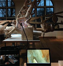
The Tropenmuseum in Amsterdam used them for their work on their 12 Bisj poles. The poles are 10 metres long and decorated using natural pigments, such as burnt ochre, which have become friable and required repair. Careful and precise cleaning and restoration was required so that dirt could be removed without causing damage, prior to overpainting. Due to the size of the poles they couldn’t be moved into the museum’s conservation studio, so instead, work was performed in a public space where the treatment was displayed on screen using a digital microscope for visitors to see.

Here is a table showing all of our current range of digital microscopes which you can select according to the required specification:
| Model |
Res. |
Magnification |
Long Working Distance |
Exchangeable Caps |
Polariser |
Metal Housing |
ESD� safe |
Additional Feat. |
Type of LEDS |
| AM4113ZT |
1.3MP |
10x-70x, 200x |
N |
N |
Y |
N |
N |
- |
8 x White |
| AM4013MZT |
1.3MP |
10x-70x, 200x |
N |
N |
Y |
Y |
Y |
- |
8 x White |
| AM4515ZT |
1.3MP |
20x-220x |
N |
Y |
Y |
N |
N |
AMR |
8 x White |
| AM7915MZT |
5MP |
10x-220x |
N |
Y |
Y |
Y |
Y |
AMR/EDOF/EDR/FLC |
8 x White |
| AM4113ZTL |
1.3MP |
20x-90x |
Y |
N |
Y |
N |
N |
- |
8 x White |
| AM4515ZTL |
1.3MP |
10x-140x |
Y |
Y |
Y |
N |
N |
AMR |
8 x White |
| AM7915MZTL |
5MP |
10x-140x |
Y |
Y |
Y |
Y |
Y |
AMR/EDOF/EDR/FLC |
8 x White |
| AM4113T-FV2W |
1.3MP |
10-70x & 200x |
N |
N |
N |
N |
N |
- |
4 x 375nm UV + 4 x white |
| AD4113T-I2V |
1.3MP |
20x-200x |
N |
Y |
N |
N |
N |
- |
4 x 390/400nm UV + 4 x 940nm IR |
| AM4115T-FUW |
1.3MP |
20x-220x |
N |
Y |
N |
N |
N |
- |
4 x 375nm UV + 4 x white |
| AM7115MT-FUW |
5MP |
20x-220x |
N |
Y |
N |
Y |
Y |
- |
4 x 375nm UV + 4 x white |